Digging more deeply into fibers and particles
Apr 29, 2020
The University of Eastern Finland is looking for a more detailed understanding of fiber reactivity and an analysis of the relationship of fibers to product properties with the Valmet Fiber Image Analyzer’s new ultra-high definition imaging and classifying capabilities.
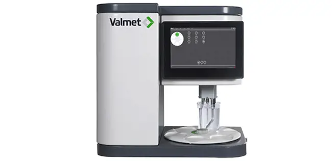
Utilizing 37 years of experience in laboratory and online fiber measurements, Valmet launched a completely redesigned optics module for the Valmet FS5 analyzer in June 2019. The ultra-high definition optics can measure more fibers with a much higher resolution to expand analysis application areas and improve measurement precision.
Since its launch in 2013, the Valmet Fiber Image Analyzer has provided precise standardized fiber morphology measurements that are possible without special training, sample preparation or laboratory facilities. The new optics now feature a wider measurement cell to allow improved shive particle measurement, a larger image area to measure more and longer fibers, and an ultra-high definition camera, providing faster fiber analysis and increased sharpness for significantly better fibril and small particle detection.
Valmet has collaborated with the University of Eastern Finland (UEF) in studying new measurement applications in the development phase of the new optics module. The target was to validate the capability of the new ultra-high definition (UHD) optics in the identification of various fines particles.
The university is now continuing to seek a more detailed understanding of fiber reactivity and to analyze the relationship of fibers with product properties.
Excelling in forest sciences
UEF’s School of Forest Sciences is an internationally renowned unit of higher education and research. The forestry and wood product-related education it provides has solid foundations in contemporary research, and UEF trains responsible experts for global forestry, land use, environment and material science issues. The Wood Materials Science MSc program, with associated research and PhD training, creates a link between wood and final products such as bio-based materials, chemicals and energy derived from renewable biological resources. The properties, performance and versatility of available raw material resources are the basis for the research, in which biomass conversion processes are being developed. Research on wood cells ranges from basic anatomical investigations related to wood tissue growth, material yield and mechanical performance to the manufacture and properties of tailored fibrous materials in paper, packaging, textiles and nanocelluloses
The applicability of the research work is broader still, meaning the results can often also be generalized for use in other areas. For example, fibrous materials are subject to manufacturing biomaterials and chemicals with a wide spectrum of end uses, ranging from biocomposites, adhesives and flocculants to rheology stabilizers and several others. They also have the potential to provide novel functionality to enhance wooden building materials for strength, decay and fire resistance.
We can visualize details far smaller than could be obtained with the device purchased in the early 2000s.
Improved small particle detection with ultra-high definition
Fast and reliable fiber characteristics analysis has already been available for decades, but the identification of small particles and different types of fine has been limited because of the optics and camera systems’ limitations. It has previously been possible to measure these different particles manually with microscopes, which is slow and tedious work, and can make it challenging to obtain a sufficient quantity of particles and fibers for reliable results.
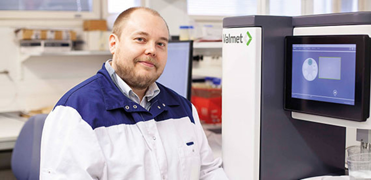
“We’re looking for small details in fibers and fines that are quite difficult to observe and require a high degree of sophistication in both optical detection and the classification of particles,” says Antti Haapala, Associate Professor at UEF. The technology in UEF’s old analyzer lacked the capabilities of “seeing” and/or identifying small particles like fiber fibrils and parenchyma cells. “We can now visualize details far smaller than could be obtained with the device purchased in the early 2000s,” Professor Haapala continues.

Fiber analysis is more than images
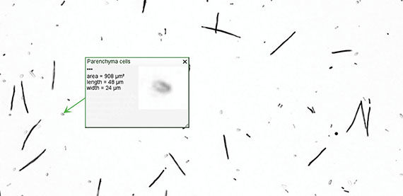
The leap from the Valmet FS5’s original high definition to ultra-high definition optics enables smaller particles to be visible. The smallest dimension of a measured particle is nearly 1 μm. This accuracy enables good identification of different fines particles from wood-based pulps. However, optical measurement capability alone is not enough. Wood- and plant-based pulps also contain other particles than fibers (tracheid cells for botanists), like vessel cells, parenchyma cells, etc. which are often very irregular. Attempting to identify these particles based on fixed parameters like dimensions, opacity, and roundness often leads to misdetections and the detection of properties that are of no interest . The Valmet FS5 has a unique built-in neural network-based feature called the Teach Tool. With this intuitive and easy to use feature, it is possible to teach the analyzer to identify different non-fiber-like particles by the user’s input of the type of particles one wishes to measure.
Professor Haapala and his team use this feature to obtain more information from their samples. “The new Valmet FS5’s features in the detection of classified particle types are being applied for the detection of different tree cell types – for example, tracheids, vessels and parenchyma. The latter two are often just considered fines,” explains Professor Haapala. After training for the analyzer has been completed, the results from the classified particles are easily available. With a typical Valmet FS5 analysis, tens or hundreds of these particles are measured, taking an average of only five minutes to complete the analysis and obtain the results from a sample. Manual analysis of these particles, in which some information may be lost or there may be insufficient particles in the microscopy sample, usually does not provide sufficient reliability. “When we’re analyzing a macerated stem wood specimen, there’s a very small amount of cut fibers and other fragments, so the Teach Tool allows us to detect these anatomical tree species-specific features,” says Professor Haapala, in describing the benefits of the Valmet FS5.
Straightforward setup
“The Valmet FS5 setup was straightforward, and everything worked out fine. The staff installing the device were very knowledgeable, and the Linux-based system of the Valmet FS5 was configured to share data on university servers running with Windows, something I was a little worried about at first. But I needn’t have been. After verification fiber checks and a briefing on user-level issues, our lab technicians began to work on our samples the same day, and several cases have already been assessed,” says Professor Haapala. At the same time, the usability of the equipment must be simple and robust, as it will occasionally be used by students. “The main differences, which also required the modification of laboratory protocols in the transition from the old fiber analysis tool, were related to automated consistency control and balancing the right quantity of particles on images for the optimal use of the Teach Tool,” Professor Haapala continues.
Helping both the academic and industrial worlds
Classification and compositional analysis of pulp fines is relevant for monitoring industrial processes such as screening and grinding, and quantifying some product properties. The morphology of fines also describes how they are generated from fibers, and the detection of fibrillar fines allows an analysis of the delamination of the fiber cell wall. The same approach has already proven effective in assessing the effects of chemical pre-treatments on fiber structure when various micro- and nanocellulose products are being prepared. An analysis of the degree of fiber deterioration is a good marker for pre-treatment efficiency and reaction kinetics, which indicates a sufficient processing time for further micronization processes.
The requirements for analysis differ based on the analyzed material: in botanical samples, it may be most important to distinguish between a cell type or assess some anatomical features in detail; in the fibrillation of celluloses, the interest is in observing how the fiber swells or delaminates during the processes. “In general, our concern is with how fiber reacts to a chemical system, or mechanical grinding or other micronization process. Fines analyses have been seen as important in many industrial applications. Versatility and the ability to customize the analysis were therefore very important for us,” Professor Haapala concludes with a happy smile.
Besides its everyday use in production facilities, the Valmet FS5 has been the analyzer chosen by many industrial research facilities and universities, providing basic data about fiber properties and new insights with its advanced features. Valmet is collaborating with their clients in research to raise the bar for measurement applications and smaller particle measurement, and for new industrial applications.
If you are interested in finding out what Valmet fiber measurements can do for you, please contact the product manager for the Valmet FS5, Tommi Niskanen (tommi.niskanen@valmet.com).
Text and photos Samar Yacoub and Antti Haapala